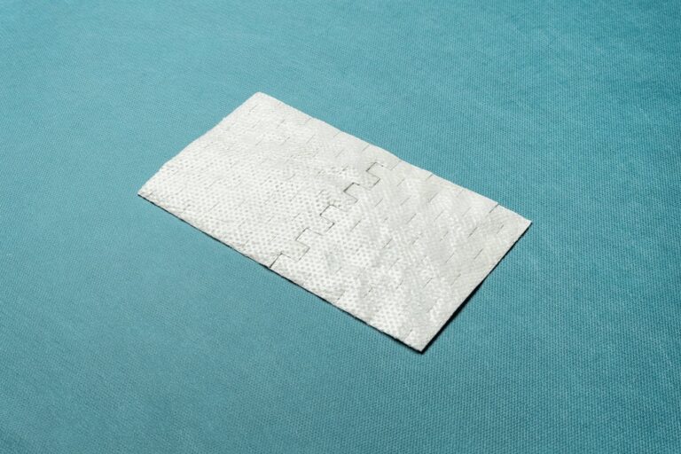
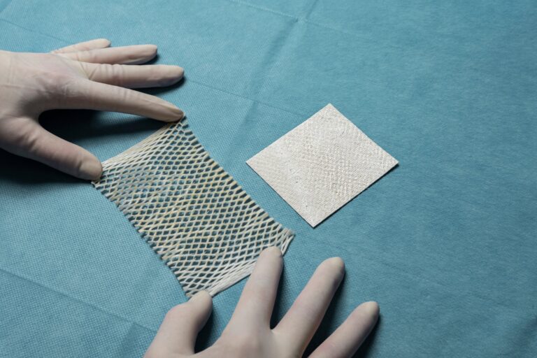
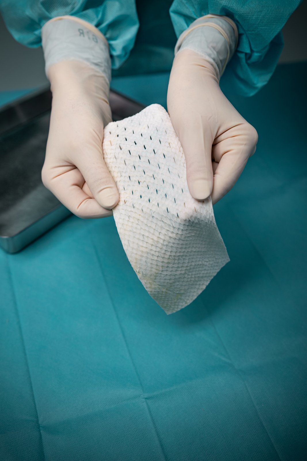
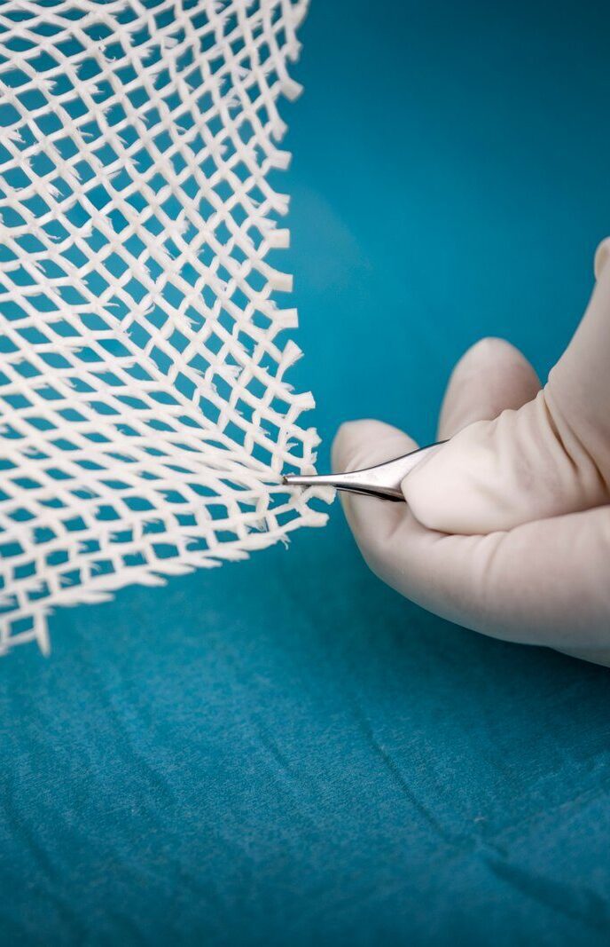
From fish skin to skin substitute

KERECIS INTACT FISH-SKIN TECHNOLOGY

Why Kerecis®?

Kerecis is intact fish skin rich in naturally occurring Omega3 polyunsaturated fatty acids. When grafted onto damaged human tissue such as a burn or a diabetic wound, the material recruits the body’s own cells and is ultimately converted into living tissue.

Compared to mammalian-based skin substitutes, Kerecis offers improved economics and clinical performance, as well as reduced disease transfer risk and no cultural constraints on usage.

Other tissue-transplant products are based on tissues of human and porcine origin. These are not ideal substitutes because heavy processing is needed to eliminate the risk of disease transmission. This harsh, anti-viral treatment removes most of the material’s natural components, making it dissimilar to human skin.

Listen to what leading authorities in the wound care field have to say about Kerecis

Dr. David Armstrong – DPM, MD, PhD

Dr. Windy Cole – DPM

Dr. Michael J. Lacqua – MD

Dr. John C. Lantis II – MD, FACS

Key technical advantages

  • No cultural or religious barriers to clinician/patient acceptance
  • Easier to use, with larger, thicker sheets (no need for multi-layer grafts)
  • Non allergenic and bio-compatible
  • No known risk of disease transfer
  • Improved infection control
  • Adjustable rate of absorption into the surrounding tissue

Fish skin has been shown to be more similar in structure to human skin than anti-viral–processed skin substitutes. Because there is no risk of disease transmission, fish skin is only minimally processed, preserving its structure and components. In a double-blind, comparative, randomized controlled clinical trial (N=162), fish skin favorably compared with mammalian skin substitutes.

Application include reconstructing the skin in burn, chronic wounds, and oral wounds, hernia repair, breast reconstruction, and dura mater reconstruction.

Other tissue-transplant products are based on tissues of human and porcine origin. Mammalian tissue carries the risk of disease transmission to humans that is nonexistent from the Atlantic cod to humans. The FDA has strict requirements on tissues from farm animals including viral inactivation methods involving treatment with detergents that remove lipids from the tissues and denature the native structure leaving behind only the most insoluble collagens. Products from human tissues like skin and dehydrated human amnion/chorion membrane call for extensive use of antibiotics to reduce bioburden. The acellular fish skin graft is not subject to this harsh treatment, leaving a more naturally intact product with its associated benefits.

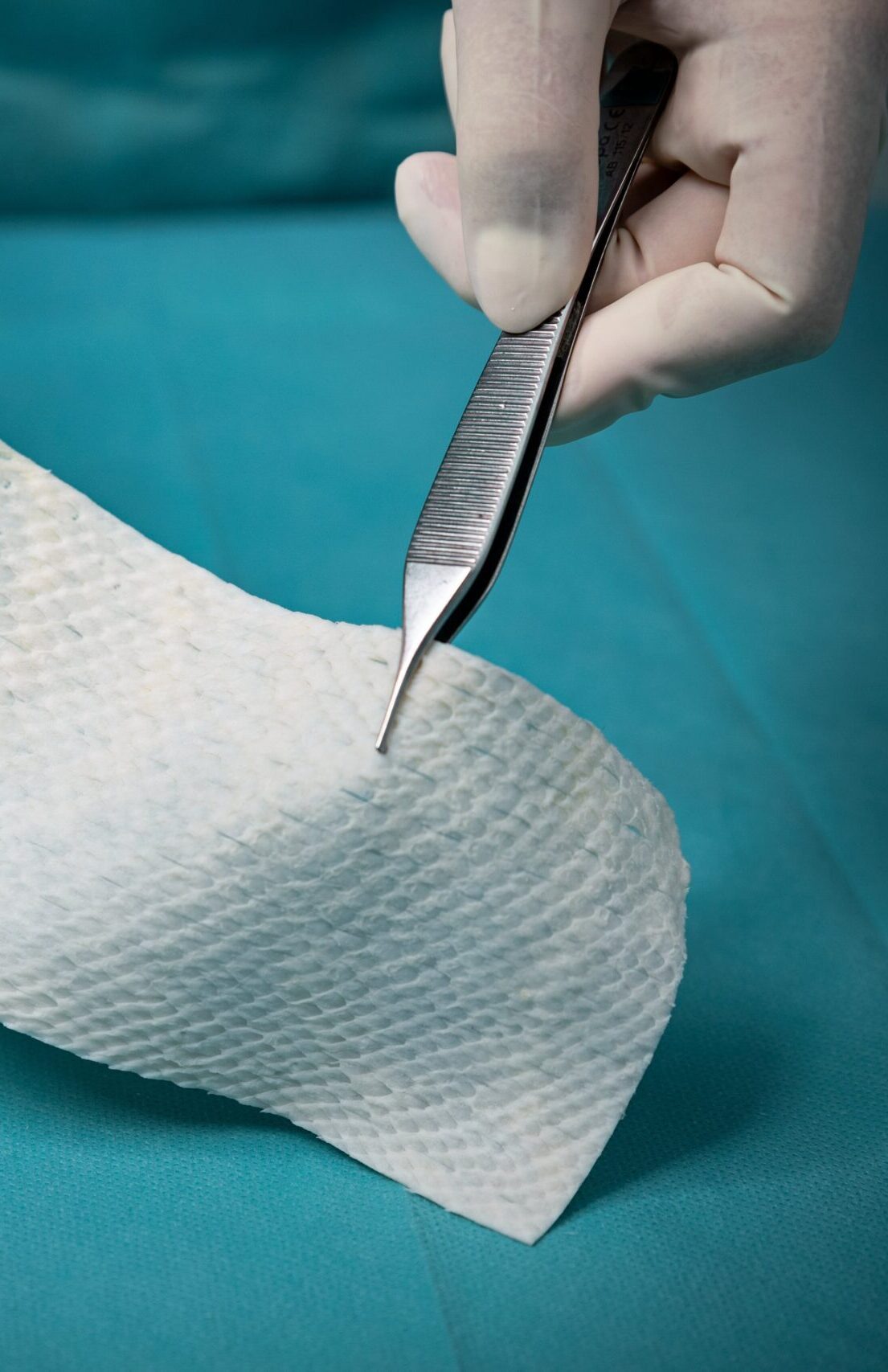
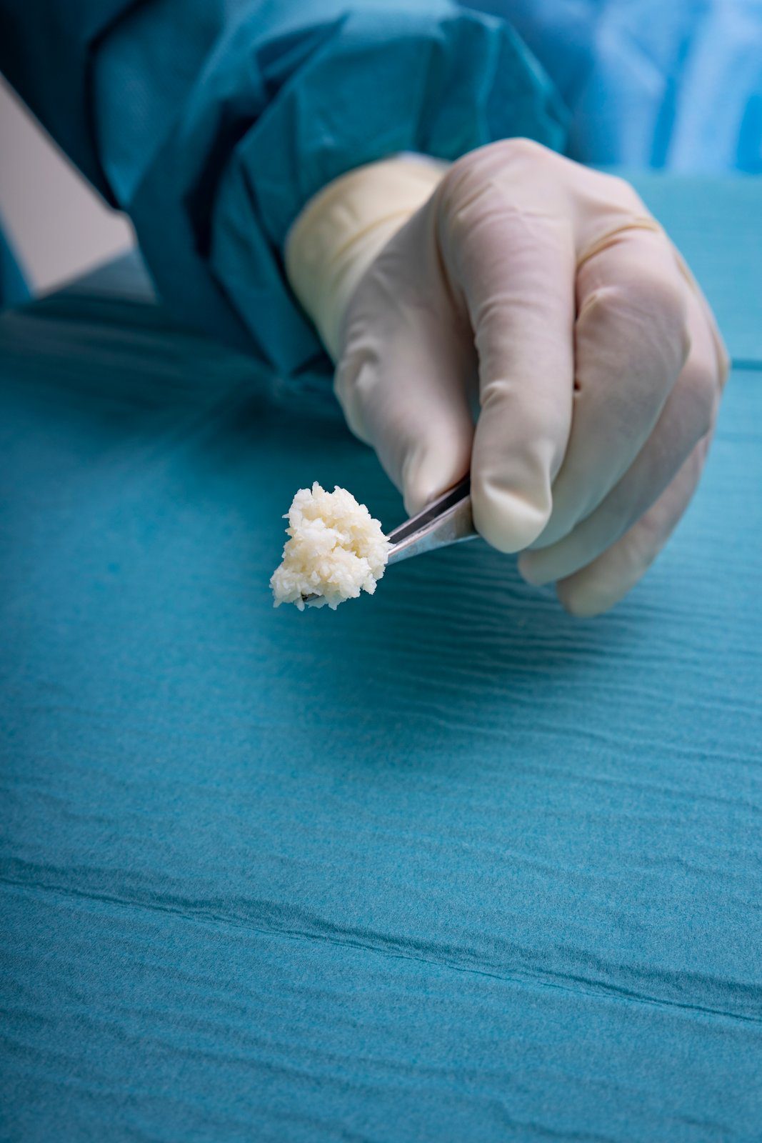
The fish are caught in the pristine waters of North Atlantic Ocean off the township of Isafjordur, on the northwest coast of Iceland. Each and every batch of raw materials is tracked to ensure product quality. The fish skin is processed using a proprietary method that preserves its structure and lipid composition.
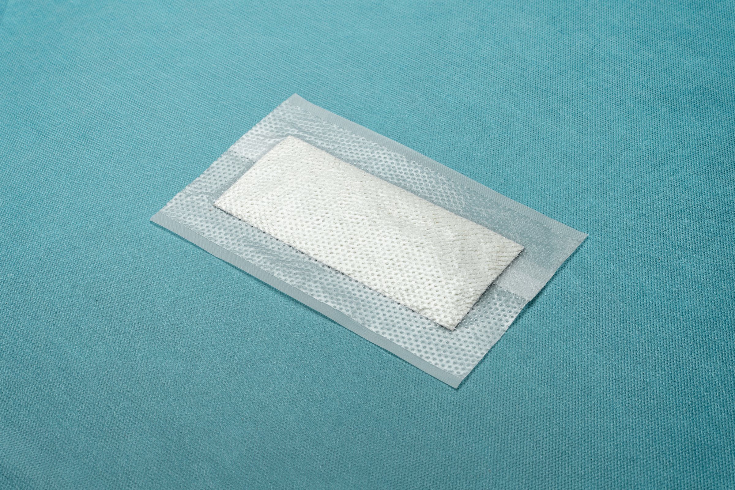
Kerecis fish skin has been approved by the FDA and European regulatory authorities for wound healing. The product is undergoing registration at multiple regulatory authorities around the world.
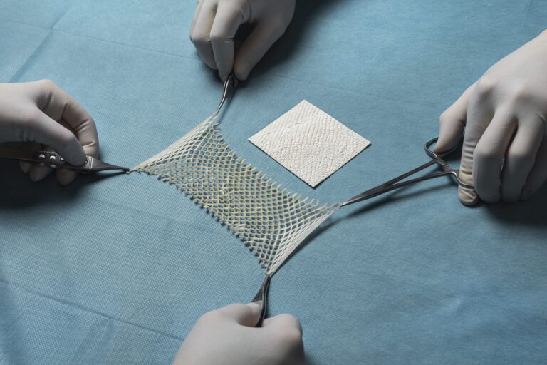
Kerecis has demonstrated its proficiency in creating lipid-containing tissue matrices from fish skin, and it has been shown that the material is safe, non-toxic and structurally sound. The company is focused on developing medical device applications with a predicated high return on investment and a low-to-medium cost of development. Kerecis focuses exclusively on tissue regeneration and maintenance, utilizing its core Omega3 fish-skin technology.

Intellectual Property

Kerecis has been awarded multiple patents protecting the core technology in the U.S. and other countries, and several more applications are pending. Kerecis is committed to building a substantial patent portfolio protecting the company´s intellectual property so that revenue from manufacturing and licensing activities can be maximized over the invention’s lifetime.

Technical Basis

The Kerecis Omega3 fish skin contains lipids and proteins that, in a concerted manner, help the body regenerate damaged tissue.

The lipids in Kerecis Omega3 include sterols, fat-soluble vitamins, and phospholipids as well as polyunsaturated fatty acids (Omega3). The health benefits of polyunsaturated fatty acids have long been recognized and their positive impact on health is on sound scientific footing [1]. These favorable effects are in large part mediated by the Omega3 (n-3) fatty acids eicosapentaenoic acid (EPA) and docosahexaenoic acid (DHA), which are found in high concentration in fish oil. One reason why wounds fail to heal is the shortage of lipids that are formed in the Golgi apparatus of the keratinocytes [2].

Kerecis fish skin

Kerecis™ Applied to the Wound

The acellular fish skin graft is derived from decellularized Icelandic codfish skin. Its protein composition closely resembles that of human skin and the porous microstructure provides a scaffold for efficient ingrowth of dermal cells and capillaries. In addition, the FDA-cleared graft is uniquely rich in the polyunsaturated Omega3 fatty acids EPA and DHA, which are known for their anti-inflammatory properties. Significant biological activity has been demonstrated [3, 4]. Kerecis Omega3 fish skin does not elicit an immune response since the major antigenic components present within cell membranes are removed during processing.

Other extra-cellular transplant products available today come from mammalian sources. Fish skin possesses many of the same features as mammalian skin, among them an appropriate surface chemistry and microstructures that facilitate cellular attachment, competent mechanical strength and biodegradation rate without undesirable by-products [6, 7, 8].

References
1. Harris WS, Mozaffarian D, Lefevre M, Toner CD, Colombo J, et al. Towards establishing dietary reference intakes for eicosapentaenoic and docosahexaenoic acids. J Nutr. 2009;139:804S-819S.
2. Feingold KR. Thematic review series: Skin lipids. The role of epidermal lipids in cutaneous Permeability barrier homeostasis. J Lipid Res. 2007;48:2531-2546.
3. Badylak SF, Freytes DO, Gilbert TW. Extracellular matrix as a biological scaffold material: Structure and function. Acta Biomater. 2009;5:1-13.
4. Reing JE, Zhang L, Myers-Irvin J, Cordero KE, Freytes DO, et al. Degradation products of extracellular matrix affect cell migration and proliferation. Tissue Eng Part A. 2009;15:605-614.
5. Lin CC, Ritch R, Lin SM, Ni MH, Chang YC, et al. A new fish-scale-derived scaffold for corneal regeneration. Eur Cell Mater. 2010;19:50-57.
6. Hawkes JW. The structure of fish skin. I. General Organization. Cell Tissue Res. 1974;149:147-158.
7. Le Guellec D, Morvan-Dubois G, Sire JY. Skin development in bony fish with particular emphasis on collagen deposition in the dermis of the zebrafish (Danio rerio). Int J Dev Biol. 2004;48:217-231.
8. Rakers S, Gebert M, Uppalapati S, Meyer W, Maderson P, et al. ‘Fish Matters’: the relevance of fish-skin biology to unvestigative dermatology. Exp Dermatol. 2010;19:313-324.
9. Image: Magnusson S, et. al. Regenerative and Antibacterial Properties of Acellular Fish Skin Grafts and Human Amnion/Chorion Membrane: Implications for Tissue Preservation in Combat Casualty Care, Military Medicine, Volume 182.1, March 2017