Advancing Wound Care with an All-in-One Product
Chronic and acute wounds can stall, become infected, or heal with excessive scarring, making timely, organized repair a clinical challenge.
In recent years, biologic materials have advanced care by modulating the wound environment, delivering bioactive components, or providing scaffolds for cells to populate.
Kerecis intact fish-skin grafts bring these elements together in a single tissue-replacement product: a skin-for-skin material that preserves the three-dimensional, layered architecture and mechanical properties of skin while retaining its natural biochemical complexity (beyond collagen alone).
This combination is designed to support cellular infiltration, vascular ingrowth, and orderly tissue remodeling — offering a practical and effective solution for managing difficult wounds.
Kerecis Mechanism of Action
Research indicates that an optimal tissue replacement product, one that not only repairs but regenerates tissue, has its cells removed from the skin without damaging or denaturing it.[1,2]
Because there is no known risk of viral disease transmission from cold-water North Atlantic cod to humans, Kerecis grafts are only minimally processed and gently decellularized using our environmentally friendly process called EnviroIntact™.
Thanks to this minimal processing, Kerecis fish-skin grafts are not damaged or denatured, and retain their molecular organization,[3] three-dimensional structure,[3] molecular content, [4,5] and mechanical properties.[4]
Together, these key features enable the fish-skin graft to promote faster and more effective wound healing by facilitating cellular ingrowth, fibroblast infiltration, blood vessel formation, granulation tissue formation, and finally, efficient incorporation into the body.

Deep Partial-Thickness Burns[6]


Burns treated with Kerecis fish-skin grafts had faster blood vessel formation and faster wound closure compared to burns treated with fetal bovine dermis (FBD).
The figure above highlights how Kerecis-treated burns had significantly more blood vessels (indicated by positive brown staining) on Day 7 following treatment.
Full-Thickness Burns[7]
Burns treated with Kerecis fish-skin grafts had similar functional and cosmetic outcomes, including pliability, pigmentation, and vascularity, as burns treated with cadaver skin.
One year after treatment with fish-skin grafts and split-thickness skin grafts (STSG), punch biopsies revealed fully regenerated skin with a natural basket-weave pattern, multiple distinct layers, and vascular structures well-distributed.
The figure above shows the epidermal-dermal junction on Kerecis-treated burns, with well-defined Rete ridges that promote healthy mechanical properties and elasticity.[8]
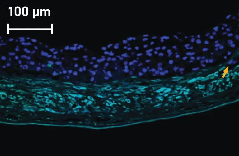
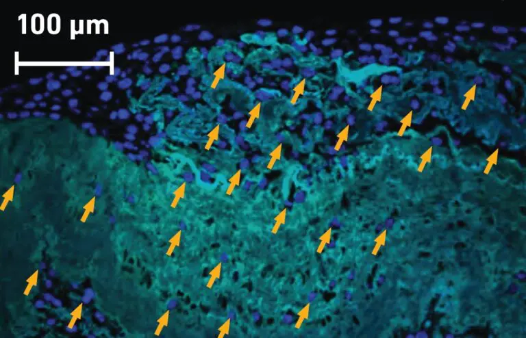
Kerecis Fish-Skin Graft has Significantly More Cell Ingrowth than Amnion / Chorion Membrane
Kerecis fish skin is homologous to human skin
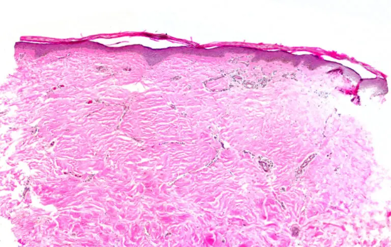
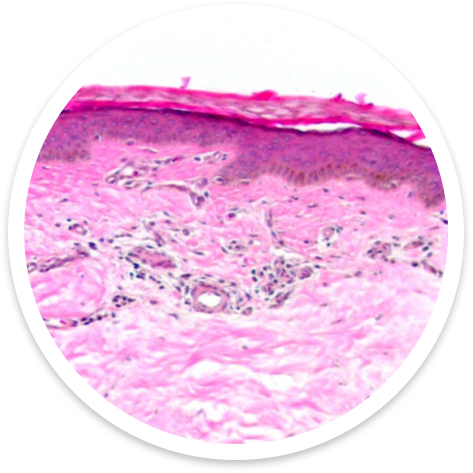
Since North Atlantic cod and humans are both vertebrates and descend from a common ancestor, the molecular organization of their skin is similar (see Figure 3).[2,3]
Both fish and human skin have an epidermis with a sheet-like morphology and underlying dermis with many of the same cell types, including Merkel cells, endothelial cells, and fibroblasts.[2]
Kerecis fish skin is also thick like human skin; our grafts’ average thickness is about 0.450 mm,3 which is slightly thicker than a typical 0.2 mm to 0.3 mm split-thickness human skin graft.9 With the similar molecular organization of fish skin and human skin, patients’ bodies are able to recognize Kerecis grafts as host tissue and can rapidly initiate wound healing.
3D Structure
Kerecis’ three-dimensional structure & porosity enables fibroblast infiltration
Our minimal processing preserves the full-thickness structure of Kerecis fish-skin grafts. Unlike cell sprays, powders, and gels on the market, Kerecis fish-skin grafts have a three-dimensional structure that provides an ideal platform for fibroblast infiltration.
Kerecis fish-skin graft and acellular human skin also have similar porosities, with pores making up 63.6% +/- 6.4% and 68.3% +/- 5.8% of space, respectively.[4,10]
This porosity is ideal for infiltration of human fibroblasts, as shown in an in vitro assay, in which notably more human fibroblasts infiltrated a Kerecis fish-skin graft than an amnion/chorion membrane product (see Figure 4).[3]
Homologous to Human Skin
Preserved Molecular Content
Kerecis’ preserved molecular content fast-tracks effective wound healing
Because Kerecis fish skin is minimally processed and gently decellularized using an osmotic gradient, the composition and structure of its extracellular matrix (ECM) remain largely intact;[4] this promotes fast and effective wound healing.
Kerecis retains its pro-healing ECM components
Our minimal processing preserves the fish skin’s ECM components, such as glycosaminoglycans, elastin, and fatty acids,[4] which are often removed during the harsher processing of mammalian tissue replacement products.11 For example, Kerecis grafts have more glycosaminoglycans, elastin, and fatty acids than bovine collagen tissue replacement products (see Figure 5).[4]
Together, these ECM components play key roles in facilitating angiogenesis, cell migration, tissue elasticity, and maintaining a bacterial barrier.[12,13]
Kerecis efficiently incorporates into the body
Kerecis fish-skin grafts are not chemically crosslinked, allowing them to easily incorporate into the wound bed.[4] For example, an in-vitro assay comparing degradation of Kerecis fish-skin grafts and fetal bovine dermis grafts found that Kerecis broke down about twice as quickly as fetal bovine dermis graft.[4]
Biodegradability of skin grafts is one of the key parameters for fast wound healing, as rapid incorporation leads to rapid release of key bioactive components.[14]

Natural Mechanical Properties
Kerecis fish-skin grafts are naturally strong and do not need artificial crosslinking to strengthen their mechanical properties
Kerecis’ minimal processing preserves the natural strength of our fish-skin grafts, including their stiffness and tensile strength.[4]
While many skin substitutes are artificially crosslinked during processing to improve their mechanical strength, there are downsides to this method. Heavily crosslinked skin substitutes can induce a foreign-body response that interferes with proper wound healing.15 Because Kerecis fish-skin grafts are naturally strong, they do not need artificial crosslinking and avoid this risk.
With their natural strength and pliability, Kerecis fish-skin grafts are also robust and easy to handle, apply, and suture. A comparison
of the mechanical properties between Kerecis and a type of bovine collagen showed that Kerecis fish-skin grafts had 56 times greater stiffness and an ultimate strength (a measure of tensile strength) that was 214 times higher.[4]

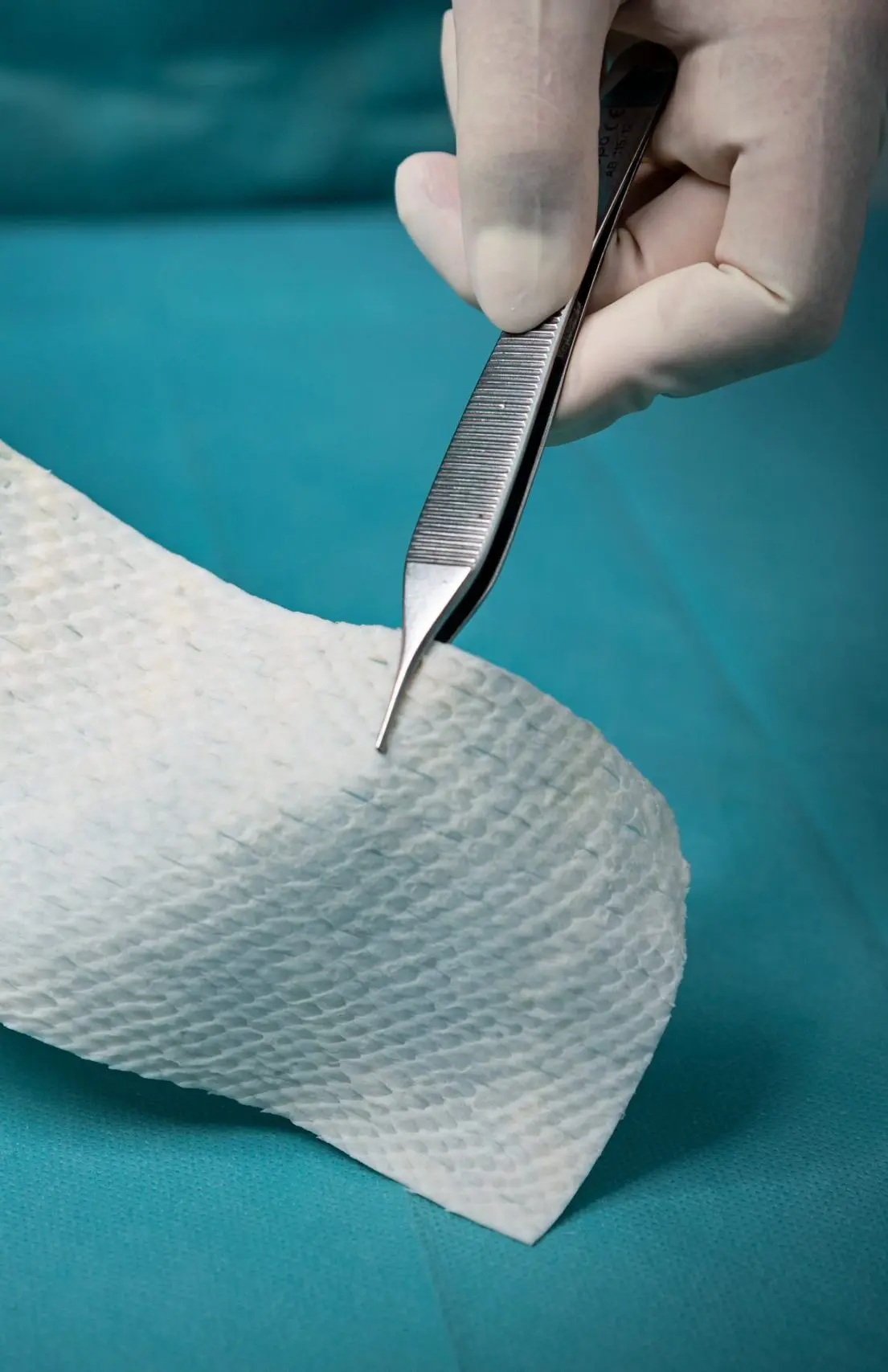
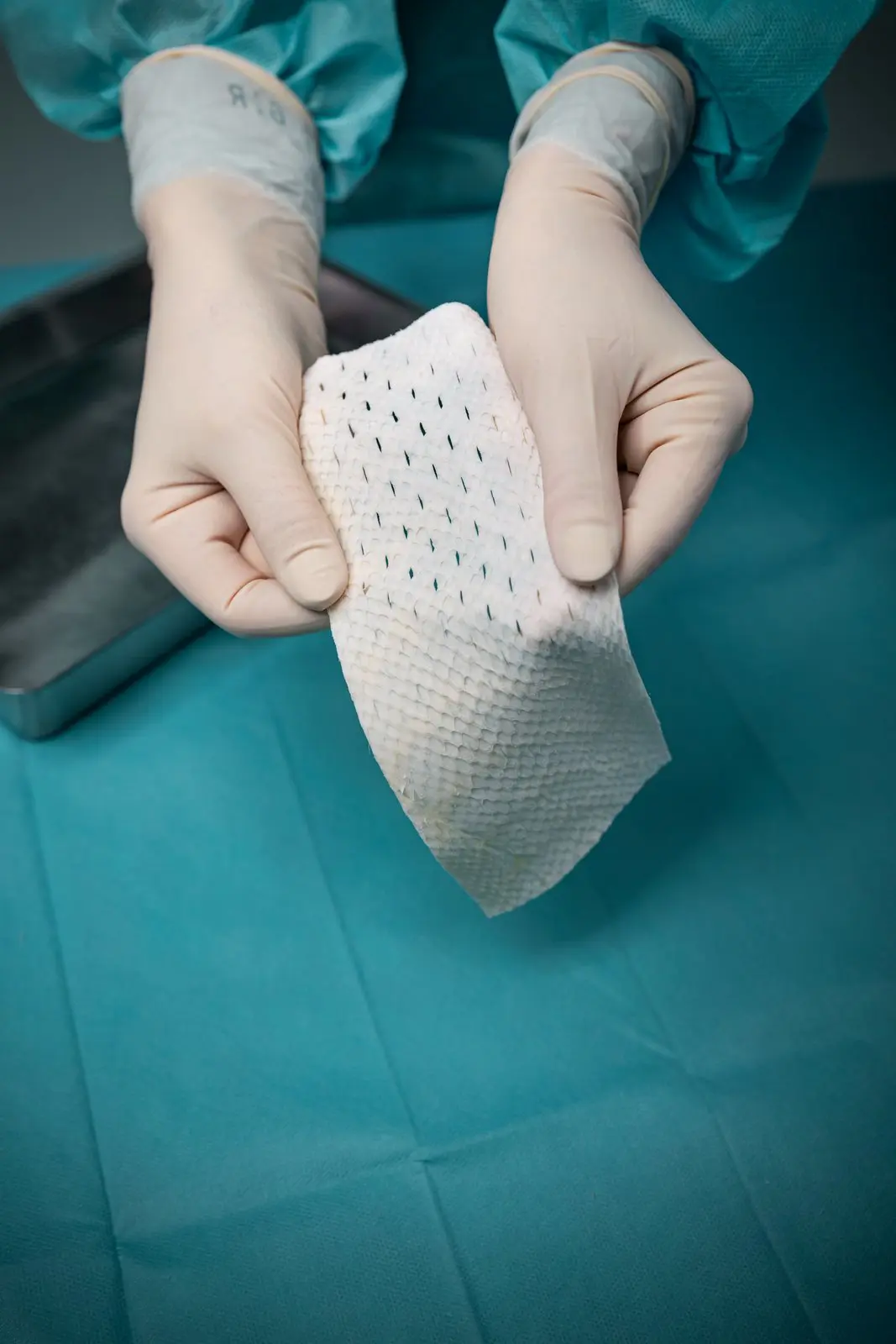
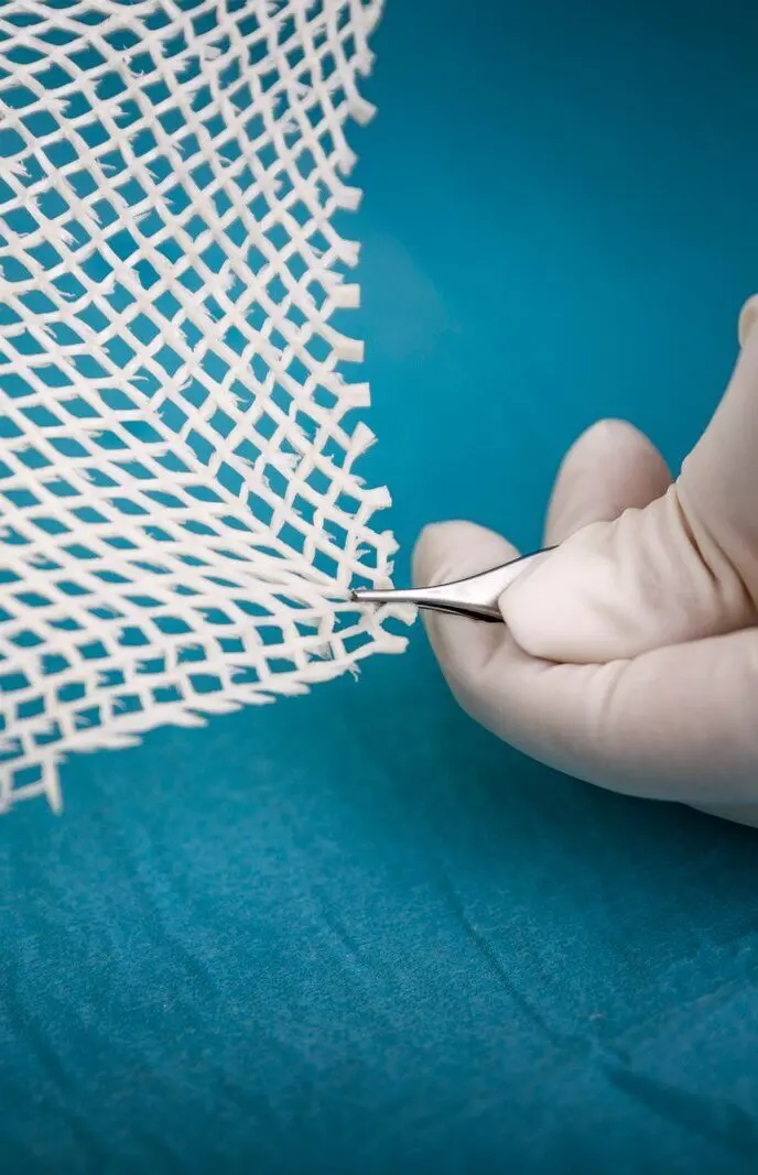
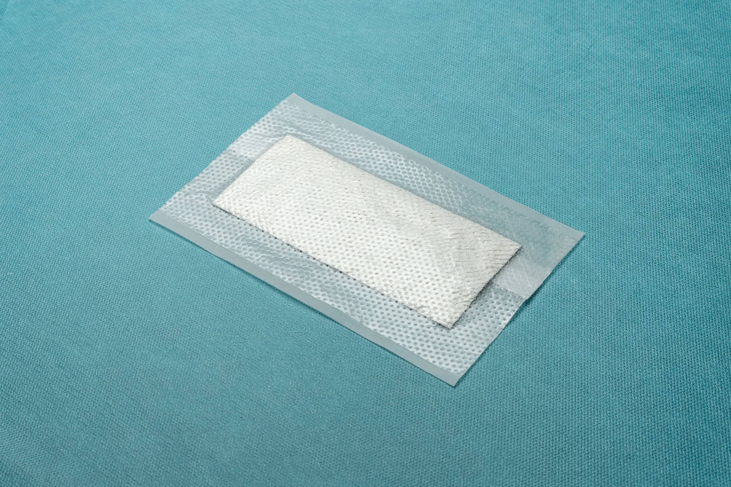
Product Configuration
Need more information?
From the town of Ísafjörður in northwest Iceland, Kerecis develops, manufactures, and distributes patented fish-skin medical devices that support soft tissue regeneration in the body, with regulatory clearance in the United States, Europe, and beyond.
Important Information
Evangelatov A, Pankov R. The Evolution of Three-Dimensional Cell Cultures Towards Unimpeded Regenerative Medicine and Tissue Engineering. In: Andrades JA, ed. Regenerative Medicine and Tissue Engineering. InTech; 2013. Accessed August 22, 2024. http://www.intechopen.com/books/ regenerative-medicine-and-tissue-engineering/theevolution-of-three…
Rakers S, Gebert M, Uppalapati S, et al. ‘Fish matters’: the relevance of fish skin biology to inves- tigative dermatology. Exp Dermatol. 2010;19(4):313- 324. doi:10.1111/j.1600-0625.2009.01059.x
Magnusson S, Baldursson BT, Kjartansson H, Rolfsson O, Sigurjonsson GF. Regenerative and Antibacterial Properties of Acellular Fish Skin Grafts and Human Amnion/Chorion Membrane: Implica- tions for Tissue Preservation in Combat Casualty Care. Mil Med. 2017;182(S1):383-388. doi:10.7205/ MILMED-D-16-001424.
Yoon J, Dogeon Yoon, Lee H, et al. Wound healingability of acellular fish skin and bovine collagen grafts for split-thickness donor sites in burn patients: Characterization of acellular grafts and clinicalapplication. Int J Biol Macromol. 2022;205:452-461.doi:10.1016/j.ijbiomac.2022.02.055
Kotronoulas A, Jónasdóttir HS, Sigurðardóttir RS,Halldórsson S, Haraldsson GG, Rolfsson Ó. Woundhealing grafts: Omega-3 fatty acid lipid content differentiates the lipid profiles of acellular Atlantic cod skin from traditional dermal substitutes. J Tissue Eng Regen Med. 2020;14(3):441-451. doi:10.1002/
term.3005
Stone R, Saathoff EC, Larson DA, et al. Accel- erated Wound Closure of Deep Partial Thickness Burns with Acellular Fish Skin Graft. Int J Mol Sci. 2021;22(4):1590. doi:10.3390/ijms22041590
Shupp J, McLawhorn M, Moffatt L. Fish Skin Compared to Cadaver Skin as a Temporary Coverage and Wound Bed Preparation for Full Thickness Burns: An Early Feasibility Trial. J Burn Care Res. 42:S124-S124. doi:doi:10.1093/jbcr/irab032.201
Blackstone BN, Malara MM, Baumann ME, McFarland KL, Supp DM, Powell HM. Laser Micropatterning Promotes Rete Ridge Formation and Enhanced Engineered Skin Strength without Increased Inflammation. Bioengineering. 2023;10(7):861. doi:10.3390/bioengineering10070861
Khan AZ, Utheim TP, Byholt M. | Tidsskrift for Den norske legeforening. Tidsskr Den Nor Legeforening.
Wang Y, Xu R, He W, et al. Three-Dimensional Histological Structures of the Human Dermis. Tissue Eng Part C Methods. 2015;21(9):932-944. doi:10.1089/ten.tec.2014.0578
Crapo PM, Gilbert TW, Badylak SF. An overview of tissue and whole organ decellularization processes. Biomaterials. 2011;32(12):3233-3243. doi:10.1016/j.
biomaterials.2011.01.057
Jiménez-Gastélum GR, Aguilar-Medina EM, Soto-Sainz E, Ramos-Payán R, Silva-Benítez EL. Antimicrobial Properties of Extracellular Matrix Scaffolds for Tissue Engineering. BioMed Res Int. 2019;2019:1-7. doi:10.1155/2019/9641456
Yue B. Biology of the Extracellular Matrix: An Overview. J Glaucoma. 2014;23:S20-S23. doi:10.1097/IJG.0000000000000108
Gregory S. Schultz GAC Lyle Moldawer, and Robert F Diegelman. Principles of Wound Healing. In: Mechanisms of Vascular Disease: A Reference Book for Vascular Specialists [Internet]. University of Adelaide Press; 2011. https://www.ncbi.nlm.nih.gov/books/NBK534261/
Badylak SF. Xenogeneic extracellular matrix as a scaffold for tissue reconstruction. Transpl Immunol. 2004;12(3-4):367-377. doi:10.1016/j.trim.2003.12.016